Monthly Breast Exam
The best time to do the breast self-exam is a few days after your period ends each month. Your breasts are less tender or swollen at this time. If you are not having periods, try to do the exam on the same day each month. Some women choose the first day of each month to help them remember.
There are two parts to a breast self-exam—looking and feeling.
Looking during a breast self-exam
The self-exam should always be done in good light. Stand or sit in front of a mirror. Place arms at your sides. Look for dimpling, puckering, or redness of the breast skin, discharge from the nipples, or changes in breast size or shape. Look for the same signs with your hands pressed tightly on your hips and then with your arm.
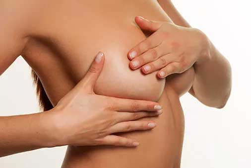
Feeling during a breast self-exam
Lie flat on your back. Place a folded towel or a pillow under your left shoulder. Place your left hand under or over your head. You also can feel for changes when you are standing. With your right hand, keeping the fingers flat and together, gently feel your left breast without pressing too hard. Use one of the three methods shown here. Then lower your right arm and do the exam on the other breast.
Choose one of these methods:
- Circle – Begin at the top of your breast and move your fingers slowly around the outside in a large circle. When you return to the top, move your hand a little closer to the nipple and make a smaller circle. Do this in smaller and smaller circles until you have examined all of the breast tissue.
- Lines – Begin in the underarm area. Slowly move your fingers down until they are below your breast. Move your fingers closer toward your nipple and go slowly back up, using the same motion. Use this up-and-down pattern all the way across your breast.
- Wedge – Begin at the outside edge of your breast. Slowly work your way in toward the nipple, doing one wedge-shaped section at a time. Do this until the entire breast area has been examined.
Ultrasound
Diagnostic Breast Ultrasound
A diagnostic ultrasound is a study performed when clinical finds are present—such as a lump, pain, or nipple discharge. This test is done in conjunction with a diagnostic mammogram. The ultrasound can differentiate cysts from solid masses. It is also a helpful tool for women who may have breast complaints but are too young for a mammogram.
Automated Breast Ultrasound (ABUS)
The ABUS system was designed specifically as a screening tool for women diagnosed with dense breast tissue. This adjunct study is similar to the 3D mammogram, but uses ultrasound to take multiple scans over sections of the breast. These fine “slices” allow the radiologist to see thru tissue often too dense to make accurate diagnoses.
Women with dense breasts are significantly better served when imaged with a combination of screening mammography and supplemental 3D automated breast ultrasound (ABUS) than when they’re imaged with screening mammography alone.
What is an Ultrasound Guided Breast Biopsy?
Click here
How is an Ultrasound Guided Breast Biopsy Done?
Click Here